An imaging test uses sound waves, radioactive particles, or magnetic fields to create an image. These images allow physician to see changes in the body that may be caused by cancer. Imaging is an essential component in the treatment of prostate cancer. Many treatments depend on the localization of cancer within the prostate to appropriately target treatment to the cancerous cells while sparing healthy tissue. Imaging is especially important for the treatment of advanced disease, as rapid detection of cancer that has spread outside the prostate (metastasis) allows patients to begin treatments earlier and increases the odds that patients will receive treatment within the optimal therapeutic window. There are many types of images used to detect and diagnose prostate cancer, the most common imaging techniques are discussed below:
Transrectal Ultrasound (TRUS):
This technique uses a small probe inserted into the rectum to send sound waves into the prostate. The echo of the sound waves off the prostate is transformed into an image by a computer. TRUS is often used to measure the size of the prostate and as a guide for biopsy and some treatments.
Computed Tomography (CT) scan:
CT scans use x-rays to create a detailed, 3D cross-sectional image of the inside of the body. CT scans may be used to pinpoint the location of a tumor within the prostate, evaluate the extent of cancer, and assess whether the disease is responding to treatment. In some cases, CT technology is used to accurately guide cancer treatment during a procedure.
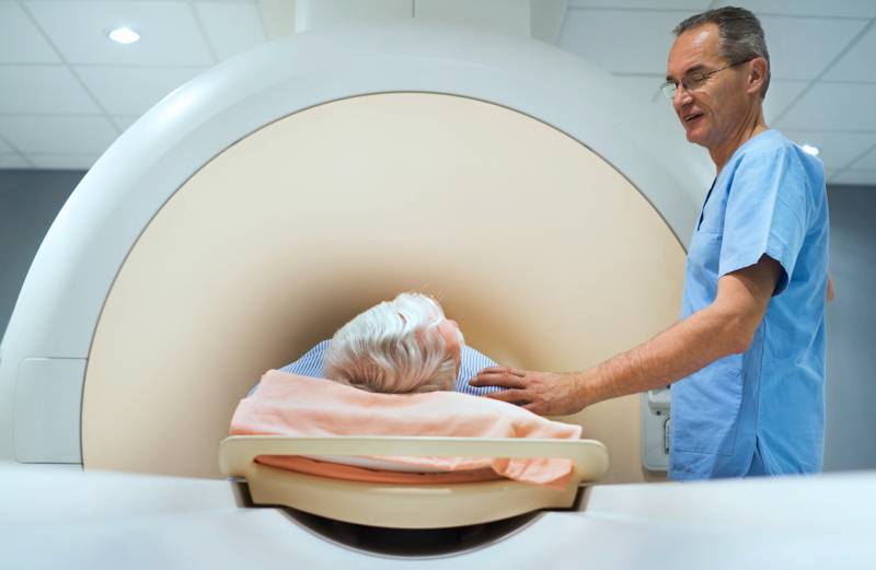
Magnetic Resonance Imaging (MRI):
MRI uses a magnetic field and radio waves to create a clear picture of the prostate. MRI can be used to locate diseased tissue within the body and to precisely pinpoint cancerous cells. MRI can also be used to see if cancer has spread outside the prostate to nearby tissues. MRI images help physicians to plan treatments such as surgery or radiation therapy.
Positron Emission Tomography (PET) imaging:
PET imaging uses a small amount of radioactive material, called a radiotracer, and a special camera to see inside the prostate. The specially designed radiotracer is injected into the patient and taken up by prostate cells, the radiotracer emits a small amount of energy which can be picked up by the PET scanner and is used to create a detailed image. This type of imaging can also identify abnormal functions in prostate cells that indicate cancer. PET Scans are usually done in combination with a CT or MRI scan.